 网站地图
网站地图

 下载:
下载:


-
量子信息技术因其优越性已经在国内外的科研领域中掀起了一番热潮。其中,利用量子体系和量子相位的演化来提高信号探测精度的量子传感逐渐成为科学家们研究的新焦点。以往人们实现量子传感主要基于超导量子干涉仪[1]或原子蒸气[2]。近几年,金刚石里的氮-空位(nitrogen vacancy, NV)色心因其在室温下具有寿命短、量子效率高且发光稳定、耐漂白性强等特点引起了科学家们的兴趣,将其发展为室温下稳定工作的单光子源[3-6]。与此同时,利用NV色心优越的光稳定性和独特的基态自旋三能级结构,人们可以通过光学可探测磁共振技术(optically detected magnetic resonance, ODMR)获取周围磁场的信息,实现高灵敏度磁场的探测。NV色心逐渐成为最近国际上最具潜力的用于磁信号探测的量子传感材料之一[7]。
NV色心作为磁场传感器的大部分工作集中于利用单个色心获得高空间分辨率的磁场探测[8-10],但利用NV色心系综则可以极大地提高磁场测量的灵敏度[11],这是由于参与相互作用的NV色心数量增多,信噪比将得到极大提升。因此,基于NV色心的磁场传感器已经被广泛运用到量子技术[12]、生物医药[13]和凝聚态物理[14]等领域当中。另一方面,利用金刚石NV色心作为增益介质实现受激辐射放大的研究已经有了较长的时间[15-17],这是因为NV色心在可见光波段具有很宽的荧光光谱,从而利用NV色心在可见光波段实现可调谐激光器拥有得天独厚的优势。近年来,在JESKE等人通过理论和实验演示NV色心在700nm处的受激辐射现象之后[18],NAIR等人通过设计一个光纤微腔在721nm处也实现了受激辐射放大[19]。但是,目前基于NV色心的激光尚未见报道,其激光阈值特性更未有研究。同时,影响激光输出阈值的参量远不止激光增益一个参量。因此,相比而言,直接采用受激辐射放大信号作为ODMR的监测信号具有诸多优点。例如:受激辐射放大信号具有与注入光相同的传输方向与偏振方向,从而使基于金刚石NV色心的远程磁场测量成为可能。本文中,作者利用含有高浓度NV色心的金刚石作为增益介质,在其零声子线(zero-phonon line, ZPL)附近观测到了受激辐射信号,并探索了其增益系数与信号光和抽运光之间的关系,为通过受激辐射放大信号实现远程磁场测量奠定基础。
-
金刚石中的NV色心由一个占据金刚石晶格位置的氮原子与一个相邻晶格位置的空缺组成。NV色心主要以两种形式存在:带负电的NV色心和电中性NV色心。如图 1a所示,NV色心具有独特的基态自旋三能级结构,处在不同基态自旋态(|0〉或|±1〉)上的粒子在被抽运到激发态上之后通过不同的途径回到基态。|0〉激发态上的粒子会直接通过辐射跃迁回到基态,而处在|±1〉激发态上的粒子更容易弛豫到中间态,在辐射一个红外的光子后无辐射跃迁回基态。因此,根据荧光信号的强度可以提取出NV色心自旋态的布居。基态自旋态|±1〉在磁场作用下会产生劈裂,从而通过扫描与|0〉到|±1〉自旋能级之间的微波共振场; 同时监测NV色心的荧光强度,便可以获得所处环境的磁场信息。此外,NV色心的吸收谱很宽,在500nm左右的激光都可以激发NV色心。而其荧光光谱在600nm~800nm的范围有一个很宽的声子边带,零声子线位于637nm附近,如图 1b所示。本实验中采用的样品是通过高温高压方法沿(100)晶面生长的高氮含量金刚石,尺寸为3.0mm×3.0mm×0.5mm。通过对该金刚石进行电子轰击和退火,大幅提高了其含有的NV色心浓度。
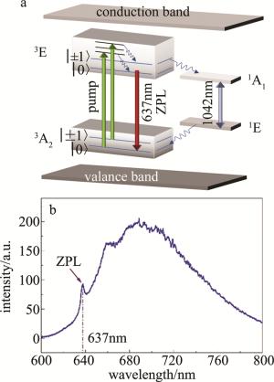
Figure 1. Energy structure and fluorescence spectra of nitrogen vacancy center
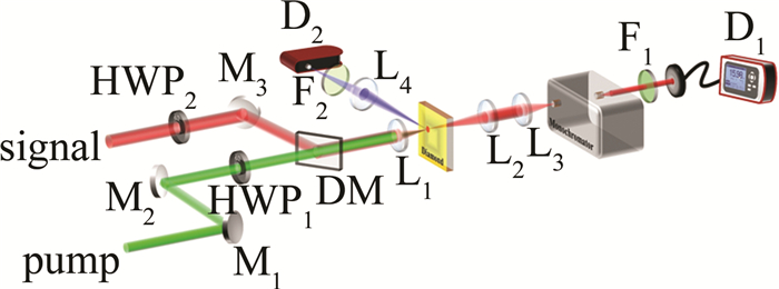
实验的光路设计如图 2所示。其中抽运光采用中心波长位于532nm的连续激光,而与NV色心的零声子线一致,中心波长为637nm的连续激光作为被放大的信号光。在激发光路部分,抽运光和信号光都为线偏振光,因此, 针对它们不同的波长选择了两个相应的半波片(half-wave-plate,HWP)HWP1和HWP2,以对抽运光和信号光的偏振角度进行调谐。抽运光和信号光通过一个600nm短通二向色镜(dichroic mirror, DM)实现合束,然后通过一个焦距为50mm的消色差透镜L1聚焦在金刚石样品的同一个位置上。经过计算高斯光束聚焦束腰,得到抽运光和信号光聚焦后光斑的直径wp=3.765μm和ws=3.381μm,wp>ws可以保证所有能产生受激辐射的NV色心都会受到抽运光的作用。在收集光路部分,为了使收集到的信号中受激辐射的信号占更大的比例,作者采用一个焦距为150mm的长焦距透镜L2来收集NV色心产生的信号。因为NV色心自发辐射所产生荧光的方向具有任意性,只有少部分的自发辐射荧光会通过L2。而受激辐射产生的光具有和信号光完全相同的频率、位相、传播方向以及偏振状态,因此理论上几乎所有的受激辐射信号都会被收集。利用光功率计和单色仪对收集到的信号光谱进行测量。在样品的侧面,放置了一个焦距为50mm的透镜L4对NV色心的自发辐射荧光进行收集,收集到的荧光信号经过580nm长通的滤波片F2滤除抽运激光,用一台光纤光谱仪D2来监测NV色心受激辐射对自发辐射荧光的抑制。
Figure 2. Experimental setup and optical path(M1~M3—mirror; HWP1, HWP2—half-wave-plate; DM—dichroic mirror; L1~L4—lens; F1, F2—filter; D1—power meter; D2—fiber spectrometer)
-
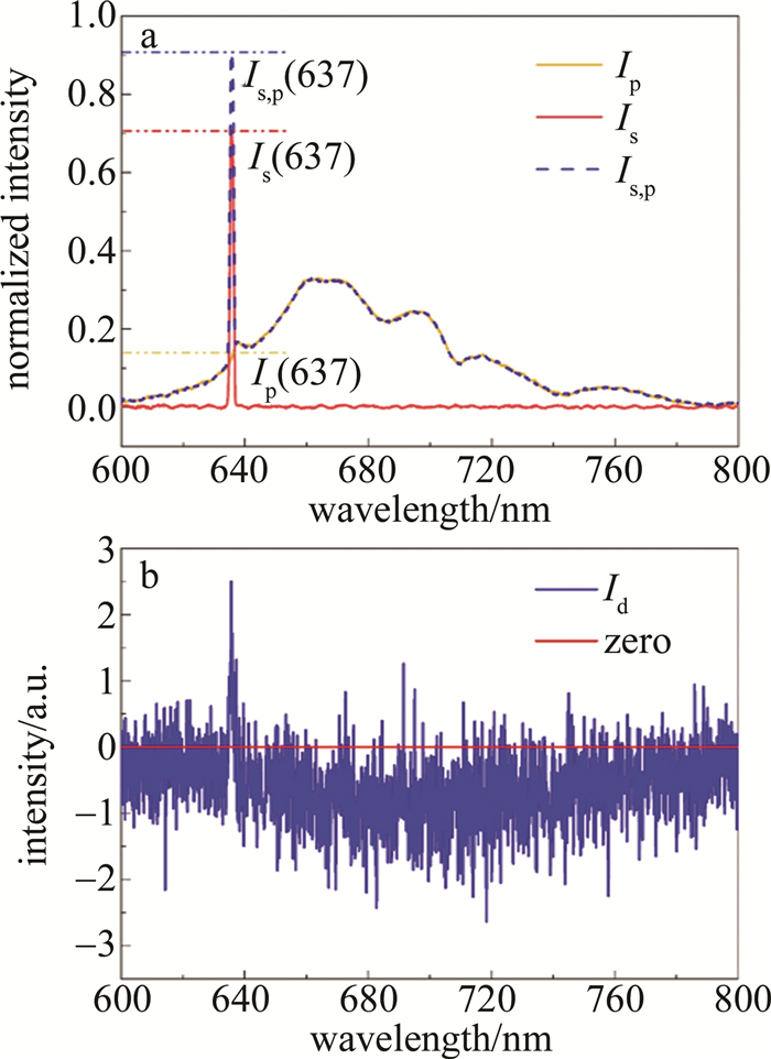
图 3a中描绘了实验过程中在不同激发条件下所收集到的典型光谱。其中仅在抽运光(信号光)作用下收集到的自发辐射光信号光强Ip(Is)用橙色(红色)实线的曲线表示,而蓝色虚线的曲线则表示抽运光和信号光共同作用下收集的既含有自发辐射光又含有受激辐射光同时还有入射光的信号光强Is, p。可以用增益系数α来表征其放大的特性:
$ \alpha=\frac{I_{\mathrm{s}, \mathrm{p}}(637)-I_{\mathrm{s}}(637)-I_{\mathrm{p}}(637)}{I_{\mathrm{s}}(637)} \times 100 \% $
(1) 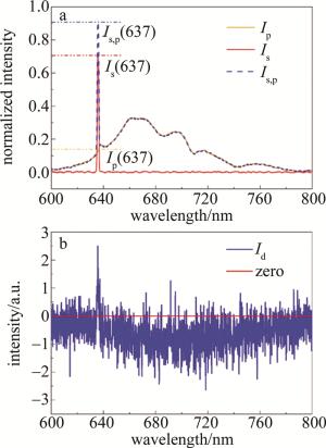
Figure 3. Acquired spectra under different conditions
式中,Ip(637), Is(637)和Is, p(637)分别表示上述3种不同激发条件下收集到的波长在637nm处的信号。在图 3a中它们的值分别为0.14,0.71和0.91,因而其增益系数的值为8.5%。而图 3b中则是通过金刚石侧面的光纤光谱仪监测到的荧光衰弱信号Id:
$ I_{\mathrm{d}}=I_{\mathrm{s}, \mathrm{p}}^{\prime}-I_{\mathrm{s}}^{\prime}-I_{\mathrm{p}}^{\prime} $
(2) 式中,Is′, Ip′和Is, p′分别表示在仅有抽运光、仅有信号光、以及抽运光和信号光同时激发金刚石这3种情况下光纤光谱仪所接收到的光强。可以看出,NV色心在信号光的作用下,其自发辐射荧光的强度降低了。因为处于NV色心激发态的粒子有一部分在信号光的作用下通过受激辐射返回到基态,因而抑制了发生自发辐射跃迁的概率,从而自发辐射荧光强度发生衰减。观察到荧光衰减的现象从侧面也反映出NV色心在信号光的作用下产生了受激辐射。此外,荧光衰减信号在637nm处观察到了一个微弱的尖峰,这应该归因于信号光有小部分散射到光纤光谱仪的方向,因而也收集到了部分受激辐射的信号。
为了尽可能地提高增益系数,首先通过旋转HWP1和HWP2来改变抽运光和信号光的偏振角度θp和θs,探究受激辐射的偏振特性对增益系数的影响,并且作者将抽运光在固定功率下激发NV色心得到荧光强度最强时的抽运光偏振方向定义为0°,而增益系数受到光的偏振角度影响可以用偏振对比度P表征:
$ P=\frac{\alpha_{\max }-\alpha_{\min }}{\alpha_{\max }+\alpha_{\min }} \times 100 \% $
(3) 在实验过程中,首先将抽运光和信号光的功率分别固定在1.25W和2.00μW,然后固定HWP2的角度,随后逐步旋转HWP1并记录每次旋转后增益系数的大小,探究增益系数与θp的关系(见图 4a),得到偏振对比度为10.2%;接着固定HWP1,再通过旋转HWP2得到增益系数与θs的关系(见图 4b),得到偏振对比度为6.9%。可以看出,增益系数会因抽运光和信号光偏振方向的改变而发生变化。这是因为沿(100)面生长的金刚石中NV色心只能有[111], [111], [111]和[111]晶向这几个固定的取向,其吸收截面和发射截面都会受到光偏振方向的影响,从而影响放大的特性。
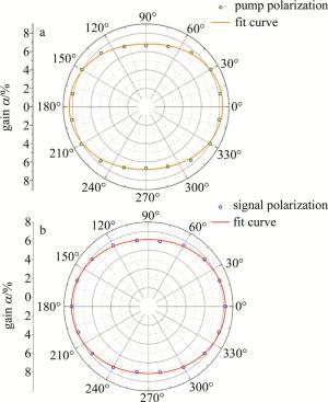
Figure 4. Influence of the polarization of pump light and signal light on the gain factor
在将抽运光和信号光的角度调整到最佳位置后,作者对增益系数与抽运光功率Pp和信号光功率Ps之间的关系展开了研究。在1.00μW~5.50μW范围内调节信号光功率,并在0.39W~2.51W的范围来调节抽运光功率优化增益系数。图 5a是增益系数受抽运光功率和信号光功率影响的等值线图。其中横坐标表示抽运光功率的大小,纵坐标表示信号光功率的大小,而旁边的颜色标尺指出了图中不同颜色所对应的增益系数的大小。图 5b中描绘了信号光为2.00μW时,增益和抽运光的关系。从图中可以很直观地看出,增益系数随抽运光功率增加而增加,这与之前初步调查的结果是一致的; 而信号光则在2.00μW左右增益系数有最大值,并在抽运光最大时(2.51W)得到了10.5%的增益系数,且呈现出增益饱和趋势。作者用一条功率饱和曲线对图 5b中的数据进行了拟合:
$ \alpha\left(P_{\mathrm{p}}\right)=\frac{\alpha_{\infty} \times P_{\mathrm{p}}}{P_{\mathrm{p}}+P_{\mathrm{sat}}} $
(4) 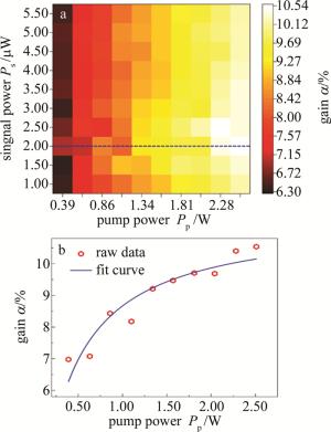
Figure 5. Influence of the power of pump light and signal light on the gain factor
式中,α∞是抽运光功率为无穷大时的饱和增益系数,Psat是饱和抽运功率。从拟合曲线中能得出α∞和Psat的值分别为11.4%±0.4%和0.32W±0.06W,因此,理论上目前这套系统的增益系数极限可以达到11.4%。
-
NV色心系综作为一个金刚石中的电子自旋缺陷,可以通过光学手段初始化和读出,因而基于NV色心的固体量子传感器在量子传感领域中备受人们的青睐。目前, 利用NV色心监测磁场的方式主要是通过收集荧光信号在微波场调控下的变化作为磁场测量的监测参量,然而受激辐射信号在传播方向上与注入信号激光方向一致,具有良好的准直性,从而为基于NV色心的远程磁场监测提供了可行性方案, 而这其中一个关键的技术需求就是要提高NV色心受激辐射的增益系数。
此次实验设计了针对NV色心零声子线的受激辐射放大系统,成功观察到NV色心受激辐射放大信号及自发辐射荧光的抑制, 并且通过优化抽运光和信号光的功率和偏振态,最终在NV色心零声子线附近取得了10.5%的增益系数,这为利用NV色心受激放大信号来远程监测磁场提供了重要的研究基础。从实验中可以发现, 这个增益系数明显受限于抽运光的功率,因而在后续的实验中,通过提高抽运光功率能够获取更大的增益系数。另外,还可以结合表面等离激元增强方法[20-21],设计表面等离激元共振增强结构来改善其受激辐射放大特性。
金刚石氮-空位色心零声子线的受激辐射放大
Amplification of stimulated radiation on the zero-phonon line of nitrogen-vacancy color centers in diamond
-
摘要: 为了解决在基于金刚石氮-空位(NV)色心的磁场高灵敏度测量中,高速获取磁场信号引起的NV色心发光强度的微小变化的技术瓶颈问题,自行设计出一套能够实现金刚石NV色心自发辐射和受激辐射信号同步测量的光学系统,并利用一个长焦距透镜收集金刚石NV色心受激辐射信号,从而尽最大可能地滤除金刚石NV色心的自发辐射信号,提高测量受激放大增益的信噪比。实验中成功观察到NV色心零声子线的受激辐射放大,分析了抽运光功率、信号光功率、抽运光偏振方向和信号光偏振方向对放大特性的影响。结果表明,通过对抽运光和信号光相关参量的优化调整,最终获得了10.5%的受激辐射增益。该研究为实现NV光放大远程磁场监测奠定了研究基础。Abstract: In order to solve the technical bottleneck of the sensitive detection of the optically detected magnetic resonance (ODMR) signal from the nitrogen vacancy (NV) color centers in the diamond by taking the advantage of the directional amplification of stimulated radiation, an optical amplification system was set up to investigate the stimulated radiation of NV color centers in diamond, where the maximum amplification efficiency reached 10.5%. Besides, it is found that the amplification efficiency is related to the pump laser power and signal laser power as well as the polarization states of the two lasers. The results show that it is promising to use the amplification of stimulated radiation to replace the fluorescence in the ODMR measurement with NV color centers in the application of remote sensing.
-
Figure 1. Energy structure and fluorescence spectra of nitrogen vacancy center
a—energy structure b—fluorescence spectra
Figure 2. Experimental setup and optical path(M1~M3—mirror; HWP1, HWP2—half-wave-plate; DM—dichroic mirror; L1~L4—lens; F1, F2—filter; D1—power meter; D2—fiber spectrometer)
Figure 3. Acquired spectra under different conditions
a—acquired by monochromator b—acquired by fiber spectrometer
Figure 4. Influence of the polarization of pump light and signal light on the gain factor
a—influenced by θp b—influenced by θs
-
[1] KITCHING J. Chip-scale atomic devices[J]. Applied Physics Review, 2018, 5(3): 031302. doi: 10.1063/1.5026238 [2] TANG J J, ZHAI Y Y, CAO L, et al. High-sensitivity operation of a single-beam atomic magnetometer for three-axis magnetic field mea-surement[J]. Optics Express, 2021, 29(10): 15641-15652. doi: 10.1364/OE.425851 [3] HUANG Sh, ZHANG W, XI Q, et al. Fabrication imperfection effect on Si/SiO2-InP micropillar cavities for 1.55μm single photon source[J]. Laser Technology, 2020, 44(5): 532-537(in Chinese) [4] CHEN Y C, GRIFFITHS B, WENG L, et al. Laser writing of individual nitrogen-vacancy defects in diamond with near-unity yield[J]. Optica, 2019, 6(5): 662-667. doi: 10.1364/OPTICA.6.000662 [5] RONG Y Y, JU Zh P, MA Q, et al. Efficient generation of nitrogen vacancy centers by laser writing close to the diamond surface with a layer of silicon nanoballs[J]. New Journal of Physics, 2020, 22(1): 013006. doi: 10.1088/1367-2630/ab6351 [6] JU Zh P, LIN J J, SHEN S, et al. Preparations and applications of single color centers in diamond[J]. Advances in Physics, 2021, X6(1): 1858721. [7] BARRY J F, SCHLOSS J M, BAUCH E, et al. Sensitivity optimization for NV-diamond magnetometry[J]. Reviews of Modern Physics, 2020, 92(1): 015004. doi: 10.1103/RevModPhys.92.015004 [8] THIEL L, WANG Z, TSCHUDIN M A, et al. Probing magnetism in 2D materials at the nanoscale with single-spin microscopy[J]. Science, 2019, 364(6444): 973-976. doi: 10.1126/science.aav6926 [9] XAVIER J, YU D Sh, JONES C, et al. Quantum nanophotonic and nanoplasmonic sensing: towards quantum optical bioscience laboratories on chip[J]. Nanophotonics, 2021, 10(5): 1387-1435. doi: 10.1515/nanoph-2020-0593 [10] TIMO W, CHRISTIAN G, FLORIAN F, et al. Determination of the three-dimensional magnetic field vector orientation with nitrogen vacany centers in diamond[J]. Nano Letters, 2020, 20(5): 2980-2985. doi: 10.1021/acs.nanolett.9b04725 [11] BIAN K, ZHENG W T, ZHENG X Zh, et al. Nanoscale electric-field imaging based on a quantum sensor and its charge-state control under ambient condition[J]. Nature Communications, 2021, 12(1): 2457. doi: 10.1038/s41467-021-22709-9 [12] SMITH J M, MEYNELL S A, JAYICH A C B, et al. Colour centre generation in diamond for quantum technologies[J]. Nanophotonics, 2019, 8(11): 1889-1906. doi: 10.1515/nanoph-2019-0196 [13] MURZIN D, MAPPS D J, LEVADA K, et al. Ultrasensitive magnetic field sensors for biomedical applications[J]. Sensors, 2020, 20(6): 1569. doi: 10.3390/s20061569 [14] CASOLA F, VAN DER SAR T, YACOBY A. Probing condensed matter physics with magnetometry based on nitrogen-vacancy centres in diamond[J]. Nature Reviews Materials, 2018, 3(1): 17088. doi: 10.1038/natrevmats.2017.88 [15] ASHFOLD M N R, GOSS J P, GREEN B L, et al. Nitrogen in diamond[J]. Chemical Reviews, 2020, 120(12): 5745-5794. doi: 10.1021/acs.chemrev.9b00518 [16] SUBEDI S D, FEDOROV V V, PEPPERS J, et al. Laser spectroscopic characterization of negatively charged nitrogen-vacancy (NV-) centers in diamond[J]. Optical Materials Express, 2019, 9(5): 2076-2087. doi: 10.1364/OME.9.002076 [17] FRACZEK E, SAVITSKI V G, DALE M, et al. Laser spectroscopy of NV- and NV0 colour centres in synthetic diamond[J]. Optical Materials Express, 2017, 7(7): 2571-2585. doi: 10.1364/OME.7.002571 [18] JESKE J, LAU D W, VIDAL X, et al. Stimulated emission from nitrogen-vacancy centres in diamond[J]. Nature Communications, 2017, 8: 14000. doi: 10.1038/ncomms14000 [19] NAIR S R, ROGERS L J, VIDAL X, et al. Amplification by stimulated emission of nitrogen-vacancy centres in a diamond-loaded fibre cavity[J]. Nanophotonics, 2020, 9(15): 4505-4518. doi: 10.1515/nanoph-2020-0305 [20] ZHAO X, DONG J, GAO W, et al. Progresses of surface enhanced fluorescence[J]. Laser Technology, 2018, 42(4): 511-520(in Chinese). [21] ARDAKANI S B, FAEZ R. Tunable spherical graphene surface plasmon amplification by stimulated emission of radiation[J]. Journal of Nanophotonics, 2019, 13(2): 026009. -

 点击查看大图

图(5)
计量
- 文章访问数: 3981
- HTML全文浏览量: 2443
- PDF下载量: 25
- 被引次数: 0
